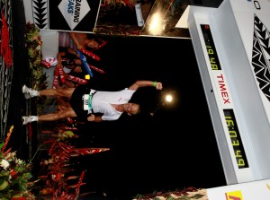
 Dr. Maroon received an athletic scholarship to Indiana University in Bloomington, Indiana where as an undergraduate, he was named a Scholastic All-American in football. Dr. Maroon has successfully maintained his personal athletic interests through participation in 9 marathons and more than 72 Olympic-distance triathlon events. However, his greatest athletic accomplishment is his participation in 8 Ironman triathlons (Hawaii – 1993, 2003, 2008, 2010, 2013; Canada – 1995; New Zealand – 1997; Germany – 2000), where he usually finishes in the top 10 of his age group. Recently, in July 2012 and 2013, he finished second and third, respectively, in his age group in the Muncie, Indiana half Ironman triathlon. In October 2013 he completed his 5th World Championship Ironman in Kona, Hawaii.
Anterior Cervical Discectomy
 Summary:
- Minimally invasive
- Small incision with typically less pain
- Home usually the next day
Background
In the spine, between all the vertebrae, there are intervertebral discs. These discs are softer than the bone of the vertebrae and they provide shock absorption and flexibility in the back. They also help keep the proper distance between the vertebrae to allow room for nerve roots to exit the spinal cord. There are two parts to the disc: a tougher outer part and an inner softer part. For this reason, the disc is often compared to a “jelly-filled donut.
An intervertebral disc can herniate, collapse or otherwise degenerate due to age, stress, strain, sudden impact or general wear and tear. If this occurs, the disc can lose its function of maintaining proper distance between the vertebrae. This can cause the nerves that exit the neck area of the spine (the cervical spine) and go to your arms to become compressed, which results in pain, numbness and possibly weakness in your arms and even legs. Neck pain and stiffness is also common and is often the first symptom to occur.
What can be done – convervative treatment
Conservative therapy, or non-surgical treatment, can be successful to relieve pain symptoms and is typically recommended as first line of treatment. Conservative therapy may include bed rest, medications, injections, physical therapy, or traction (i.e. the use of mechanical force to put tension on the displaced vertebrae to put them back in place). A “nerve block injection” to the specific nerve(s) in the neck that likely are compressed and cause the pain can both relieve symptoms and determine if the injected nerve indeed is the nerve that is compressed and causes the pain. A nerve root injection may be necessary prior to surgery, to better determine if the ACDF surgery will help you.
ACDF surgery – Anterior Cervical Discectomy and Fusion
Once diagnosed with a cervical herniated or collapsed disc and having failed conservative treatment, an Anterior Cervical Microdiscectomy and Fusion surgery is often recommended to decompress the nerves and spinal cord.
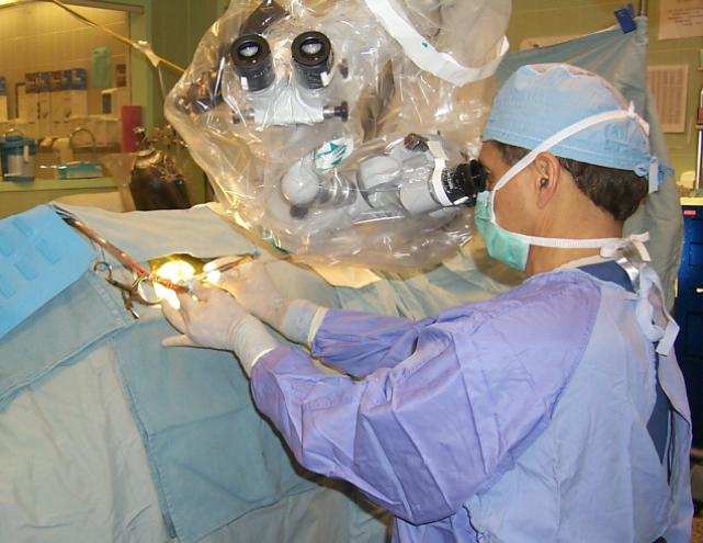
 The Anterior Cervical Microdiscectomy and Fusion (ACDF) surgery is designed to remove the material that is compressing the nerve; this can be the herniated disc material, bony compression due to arthritis or bone spurs from the vertebrae that stick into the nerve. The removal of the material is then followed by a fusion of the directly surrounding vertebrae. This fusion will stabilized the area and hold the bones in place with adequate distance to allow healing and reduce the pain associated with movement in that part of the neck.
The ACDF procedure both removes the nerve pressure and stabilizes the spine, which typically results in relieve of the arm symptoms. For the ACDF procedure, Dr. Maroon uses minimally invasive techniques, which allows for reduced tissue trauma and hence faster healing times. In most cases, the patient is released from the hospital the next day and is quickly able to return to many of the daily activities. There are generally no behavioral restrictions 1 to 3 months post-surgery.
How is the ACDF Surgery is Done?
The ACDF surgery is performed through a small incision (1 to 2 inches in length), made usually on the right side of the neck, about at the level of the Adam’s apple (see video below). Due to the minimally invasive nature of the surgery, in order to get the front area of the spinal column, the muscles of the neck are typically moved out of the way instead of being cut. The disc material and/or bone spurs are being removed so that any pressure on the nerves or spinal cord itself from disc or bony material will be alleviated. Lastly, the (former) disc space is reconstructed with a spacer. This reconstruction in the neck area is called anterior cervical fusion. The ACDF surgery usually takes about one to two hours.
There are two methods of cervical fusion: with plate and without plate.
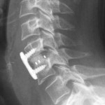
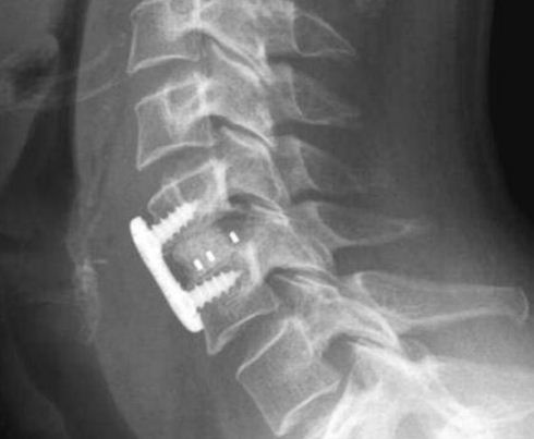
For fusion with plate, a spacer is placed in the disc space, and a metal plate is placed over the front of the disc space to hold the spacer in place and to prevent movement between these vertebrae. The inserted spacer promotes bone growth and eventually, the two adjacent vertebrae will grow together and provide the space and stability necessary. Until this process is completed, the plate will have that function. The plate is made of very strong, non-magnetic metal (which will not set off a metal detector) and is held in place with two small screws (see x-ray photo). The plate will not be removed after the bone fusion process of the body is complete unless necessary for medical reasons.
For fusion without plate, see Cervical Fusion without a Plate.
How Successful is this Surgery?
The success rate of this type of surgery is 80 to 90%. However, all procedures have risks. Potential anesthetic complications will be reviewed with you by the anesthesiologist on the day of surgery. Following the surgery, minor discomfort resulting from the incision is common, but temporary. You may also have some pain with swallowing. If such pain occurs, it can be relieved with the use of mild pain medication. Typically, you will not need to wear a cervical brace after surgery.
Tri-State Neurosurgical Associates-UPMC
Office Addresses:
Administrative Oakland Office
Presbyterian University Hospital
Department of Neurosurgery
Suite 5C
200 Lothrop Street
Pittsburgh, PA 15213
Phone: 1-888-234-4357
© 2013 Tri-State Neurosurgical Associates – UPMC