
 Dr. Maroon received an athletic scholarship to Indiana University in Bloomington, Indiana where as an undergraduate, he was named a Scholastic All-American in football. Dr. Maroon has successfully maintained his personal athletic interests through participation in 9 marathons and more than 72 Olympic-distance triathlon events. However, his greatest athletic accomplishment is his participation in 8 Ironman triathlons (Hawaii – 1993, 2003, 2008, 2010, 2013; Canada – 1995; New Zealand – 1997; Germany – 2000), where he usually finishes in the top 10 of his age group. Recently, in July 2012 and 2013, he finished second and third, respectively, in his age group in the Muncie, Indiana half Ironman triathlon. In October 2013 he completed his 5th World Championship Ironman in Kona, Hawaii.
Orbital Tumor Surgery
 The orbital part of the skull refers to the bony socket that contains and protects the eyeball. The orbit has a large opening to the front where the eye lens comes out, and has several small openings in the back that allow for the optic nerve, blood vessels and other nerves to connect the eye to the brain. The orbital region includes the surrounding bone and the muscles that control the eye, as well as nerves and blood vessels. While orbital tumors arise most often from nerves, they can arise from any of the orbital structures.
Types of tumors
Like most other brain tumors, orbital tumors usually are bening, although malignant tumors do occur. Orbital tumors can arise from cells in the sheaths that cover all nerves, called Schwann cells and the tumors then are called neurofibromas and schwannomas. The only exception to this is the optic nerve, which doesn’t have Schwann cells, so this type of tumor cannot occur on the optic nerve. However, glial cells are cells in the optic nerve and in the brain that can be affected by tumors called optic gliomas. Another type of tumor is the orbital meningioma, which comes from the meninges, the protective covering that surrounds the brain and optic nerves. These tumors may arise within the orbit, or may develop outside the orbit and grow to include it. Other tumors can arise from the bone or the skin around the eye. While most of the orbital tumors arise from the orbital area, some tumors may spread to the orbit from other parts of the body (metastasize).
Symptoms and diagnosis
The most common symptom of an orbital tumor is the protrusion of the eye. Tumors in this region also often cause pain or a feeling of pressure in the eye socket. Orbital tumors that involve neural structures typically cause significant visual problems, such as double vision and vision loss, whereas tumors that do not involve neural structures usually cause milder visual problems (by compressing the optic nerves).
Imaging studies are key to the accurate diagnosis of orbital tumors. While both Magnetic Resonance Imaging (MRI) and Computed Tomography (CT) scans can be used, MRI’s are usually preferred due to the clear images they provide of the orbital structures. Before the MRI or CT scan, the patient will be given a “contrast” agent through an IV that helps the neurosurgeon visualize the normal tissue versus the tumor. Also, in some cases a biopsy of the tumor will be taken in order to examine the tissue under a microscope and to provide a definitive diagnosis.
Is treatment necessary?
Since most neurological tumors in this region are benign, if a tumor is small and does not cause symptoms, it may be observed without treatment. However, if the tumor causes disfiguring protrusion of the eye or begins to cause visual problems, surgical removal is the best treatment option. For benign tumors, surgery usually will provide a cure. For rare malignant tumors, surgery may have to be followed by radiation and/or chemotherapy.
The surgery
In some cases, you and your neurosurgeon will decide that orbital tumor surgery is the best option for you. Before the surgery, a thorough explanation of the surgical procedure including risks, benefits, and alternatives will always be provided as it pertains to your specific case. Also, your current level of vision and eye muscle function will be thoroughly assessed and documented.
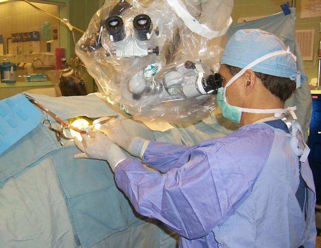
The surgery itself can consist of a craniotomy, where the tumor is being approached from the outside through the skull, or with the Endoscopic Endonasal Approach (EEA) through the nose, with no incision. The best method depends on the type and exact location of the tumor.
The biggest concern during or after surgery is that vision can be negatively affected due to trauma during the surgery or bleeding or swelling after the surgery. Vision will be closely assessed and monitored several times right after surgery. Due to the fact that the orbital area is crowded with tiny, yet very important structures such as nerves and bloos vessels, other side effects of orbital surgery to the face or brain are also a risk. Of course, your individual risks will be discussed in detail with you
Why Dr. Maroon at UPMC?
Dr. Joseph C. Maroon has been recognized as world leaders in the surgical treatment of orbital tumors. He has presented at numerous national and international neurosurgical and ophthalmologic conferences and has contributed over 17 book chapters and many scientific articles to the medical literature specifically about surgical approaches to orbital tumors. But most importantly, through many years of experience and many hours in the anatomy lab, Dr. Maroon has developed and perfected many of the modern surgical techniques used to treat orbital tumors. Orbital tumor surgery is a routine surgery for Dr. Maroon and his team at UPMC, and he is the preferred referral surgeon for physicians from all over the world. At UPMC, Dr. Maroon has access to state-of-the-art equipment and diagnostic tools that are used to diagnosis and treat tumors of the orbits as safely, efficiently and minimally invasively as possible.
Tri-State Neurosurgical Associates-UPMC
Administrative Oakland Office Address:
Presbyterian University Hospital
Department of Neurosurgery
Suite 5C
200 Lothrop Street
Pittsburgh, PA 15213
Phone: 1-888-234-4357
© 2013 Tri-State Neurosurgical Associates – UPMC