 Dr. Maroon received an athletic scholarship to Indiana University in Bloomington, Indiana where as an undergraduate, he was named a Scholastic All-American in football. Dr. Maroon has successfully maintained his personal athletic interests through participation in 9 marathons and more than 72 Olympic-distance triathlon events. However, his greatest athletic accomplishment is his participation in 8 Ironman triathlons (Hawaii – 1993, 2003, 2008, 2010, 2013; Canada – 1995; New Zealand – 1997; Germany – 2000), where he usually finishes in the top 10 of his age group. Recently, in July 2012 and 2013, he finished second and third, respectively, in his age group in the Muncie, Indiana half Ironman triathlon. In October 2013 he completed his 5th World Championship Ironman in Kona, Hawaii.
Cranial Anatomy
We will briefly describe the following parts of the anatomy in the head:
- Brain Coverings – describes how the brain in embedded in the skull
- Brain Anatomy – describes different parts and functions of the brain
- The four brain lobes – describes the different parts and functions of the cerebral cortex, the most sophisticated part of the brain.
Brain Coverings
The human brain is generally well protected from outside injury. It is firmly surrounded by three layers of membranes, encased in a rigid skull (the cranium), and covered by a muscular scalp. Each of these barriers to the brain is important, because brain tissue is fragile and can be unforgiving if injured.
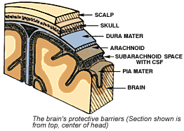
The three layers of membranes (the meninges) that cover the brain and the spine are the:
- Dura mater, a tough and fibrous protective membrane that lines the skull from the inside
- Arachnoid mater, a thin membrane which provides cushioning
- Pia mater, a very fine membrane, and the only one directly lining the surface of the brain by following all the grooves. It provides nourishment to the brain through its many fine blood vessles.
The space between the arachnoid mater and the pia mater is filled with a fluid that provides nutrients as well as shock-absorption to the brain: the cerebrospinal fluid (CSF). The CSF also surrounds the spinal cord and fills open spaces (ventricles) inside the brain. The amount of CSF that circulates around the brain normally stays the same. The CSF helps to maintain a constant pressure inside the skull, known as intracranial pressure (ICP).
Brain Anatomy
We may think of the brain as one organ, but it actually consists of many different structures and areas, all of which have specific functions for your body and mind. Because of the large number of different structures, it is customary to describe functions of brain areas that summarize the function of all the different structures included. We can divide the brain into three main parts: the forebrain, midbrain and hindbrain.
The hind brain and mid brain (together: the brain stem)
The hind brain has as main structures the pons, medulla and cerebellum (small brain), and the mid brain connects the hind brain with the forebrain. The hind brain and the mid brain together form the “brain stem”. The brain stem is located in the very center of the brain, and is a direct extension of the spinal cord. This is the oldest structure of the brain, and it is responsible for the most basic functions necessary to live, including maintaining consciousness and regulating heart rate, breathing and the sleep cycle. Also, the nerve connections between the motor system and sensory systems pass through the brain stem. The brain stem functions as main circuit for all brain activity and anchors the brain to the other part of the central nervous system and the spinal cord.
The forebrain
The forebrain is the largest and most sophisticated part of the brain. It has two parts to it: the diencephalon and the cerebrum.
Diencephalon
The diencephalon is located under the cerebrum, and above the brain stem. The most important structures here are the thalamus and the hypothalamus.
The diencephalon functions as a relay system that receives and filters incoming sensory information from the body and then relays it on to the right parts of the brain where it can get processed (mainly the cerebral cortex, but also to the cerebellum or brainstem). It also helps to connects structures of the endocrine system (hormone system) with the nervous system, and it works together with the limbic system in creating and managing emotions and memories. The diencephalon is also responsible for autonomic function control.
Cerebrum (also called telencephalon)
The cerebrum is the part on the very outside of the brain, which is also the largest part of the brain. The outmost layer of the cerebrum is called the cerebral cortex. The cerebrum is divided into two sides, the left and right cerebral hemispheres. The hemispheres are connected and communicate through a thick bundle of nerves, the corpus callosum.
The cerebrum controls most of the body’s thought and sensory processes. More specifically, this part of the brain determines personality and intelligence and is responsible for abstract tasks like planning and organizing. It also plays an important role in interpreting sensations such as touch and smells. See the part about brain lobes for more specific information.
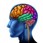
The four brain lobes
Both cerebral hemispheres can be divided into four main areas, or lobes, each of which is responsible for certain sensory and cognitive functions.
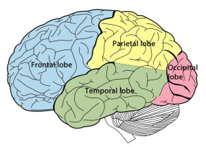 Frontal lobe
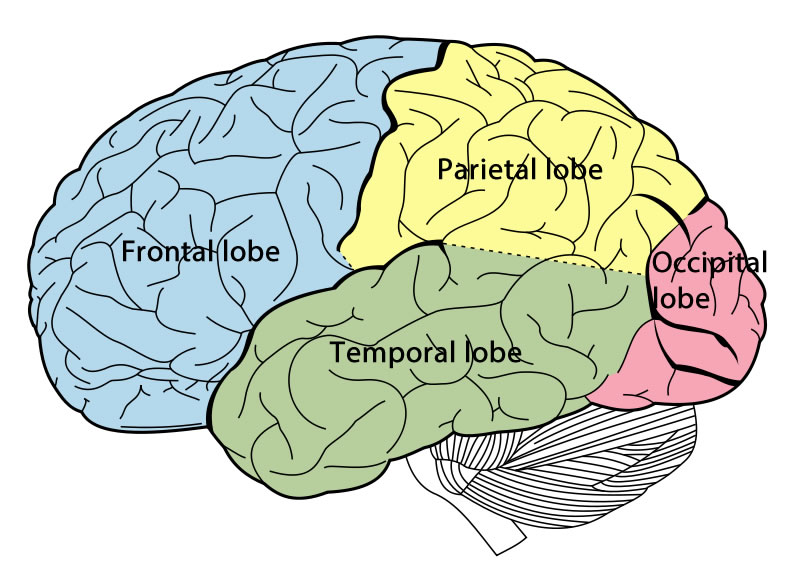
The frontal lobe is associated with complex thought and reasoning, expressive language, emotions and personality. This lobe also includes a part called the motor cortex, an area which received information from the other lobes and uses this information to purposefully carry our body movement. The frontal lobes are the most uniquely human of all the brain structures. Damage can result in mood changes, attention problems, socialization or personality changes, and decreased inhibition resulting in changes in sexual habits and increased risk-taking.
Parietal lobe
The parietal lobe helps integrating sensory information from various senses, such as pressure, touch and pain, and it is largely responsible for the visuo-spacial orientation and processing. Damage can result in problems with verbal memory, other problems with language, and difficulty to control eye gaze.
Temporal lobe
The temporal lobe is responsible for the senses of smell and sound, as well as for the processing of complex stimuli like faces and scenes. This lobe includes the primary auditory cortex, which is important for interpreting sounds and the language we hear. It also includes the hippocampus, an important structure for the consolidation of information from short-term memory to long-term memory. Damage can lead to problems with language skills, speech perception and memory.
Occipital lobe
The occipital lobe is responsible for interpreting visual stimuli and information. This lobe includes the primary visual cortex, which receives and interprets information directly from the eyes. Damage to this lobe can cause visual problems such as difficulty recognizing objects, colors and words. Damage can also produce hallucinations.
Tri-State Neurosurgical Associates-UPMC
Administrative Oakland Office Address:
Presbyterian University Hospital
Department of Neurosurgery
Suite 5C
200 Lothrop Street
Pittsburgh, PA 15213
Phone: 1-888-234-4357
© 2013 Tri-State Neurosurgical Associates – UPMC